The Hidden Epidemic of Low Back Pain
Low back pain is the leading cause of disability worldwide. In 2020 alone, it affected approximately 619 million people globally, a number projected to surge to 843 million by the year 2050. The economic and societal burden of this epidemic is staggering; it is a primary driver of physician visits, workplace absences, long-term disability claims, and prescriptions for opioids.
Yet, despite the massive toll it takes on the economy and workforce productivity, doctors frequently hit a wall when trying to diagnose it. Current standard diagnostic tools, such as X-rays, CT scans, and MRIs, are great at spotting large scale anatomical problems like a fully herniated disc. However, they often completely miss the subtle, early-stage molecular breakdowns happening inside the spine. As a result, many patients suffer from debilitating pain while their MRI results appear perfectly "normal".
Looking Closer: The Power of Advanced Laser Microscopy
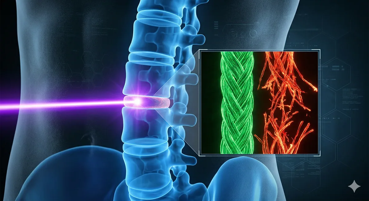
A recent master's dissertation by Sasha Dawn MacArthur at Saint Mary's University may hold the key to solving this diagnostic blind spot. The research focuses on the intervertebral discs (IVDs), which is the natural "shock absorbers" located between the bones of our spine. These discs rely heavily on a strong outer ring made of collagen, a tough, rope like protein. When a disc degenerates, this highly organized collagen network begins to fray and disorganize at a microscopic level long before the disc actually bulges or tears.
To see this invisible damage, MacArthur’s research utilizes a cutting-edge technique called Polarization-Resolved Second Harmonic Generation (PSHG) microscopy.
What is PSHG? In simple terms, it is a highly specialized laser imaging technique. It fires ultra-fast pulses of light at tissues. When these specific light pulses hit highly ordered structures like collagen, they bounce back a new signal at exactly half the original wavelength. By analyzing how this light interacts with the tissue, scientists can calculate exact mathematical parameters of "disorder" in the collagen without ever having to use chemical dyes or invasive tissue labels.
The Findings: Catching Damage Before It's Too Late
The study analyzed human spinal tissue from patients who underwent surgery for lumbar disc herniation, comparing different grades of disc degeneration. MacArthur tested several variations of this laser technique and found that one specific method, called "PIPO-SHG", was highly robust and reliable at mapping out the molecular chaos in degenerating spinal discs.
The findings were revealing: the laser microscopy successfully detected progressive molecular and fibril disorganization in the collagen of degenerating discs. Crucially, the data suggested that these structural alterations in the collagen happen prior to the onset of visible bone and endplate diseases (known as Modic changes) that doctors usually look for on MRIs.
Economic Impact and Industrial Opportunities
The transition of this technology from the laboratory to the commercial medical device industry could be transformative. Because PSHG microscopy can quantify exactly how "disordered" a spinal disc's collagen has become, it creates a massive industrial opportunity to manufacture next-generation diagnostic imaging systems.
From an economic standpoint, the current chronic management of low back pain relies on non-specific treatments that offer limited long-term relief. A commercial medical device utilizing PSHG could allow doctors to catch spinal degeneration at its true molecular onset. This means that instead of paying for years of ineffective pain management, opioid prescriptions, and eventual emergency surgeries, healthcare systems could intervene early with targeted physical therapies, regenerative medicine, or preventative care.
While PSHG is currently a sophisticated laboratory tool requiring complex laser setups and sometimes time-intensive data processing, this research lays the precise groundwork for biomedical engineering companies to optimize and commercialize the technology for clinical and preclinical use. If capitalized upon, spotting the "invisible" origins of back pain could save economies billions of dollars in lost productivity and radically shift the landscape of musculoskeletal healthcare.